Meet your Executive Board: Scott Dudis
 Thursday, April 11, 2013 at 12:00PM
 This post is part of a series made to introduce you to your elected board members, and also to share a little bit about what SAVMA is working on on a national level. The officers are a group of students that are former SAVMA Delegates and were elected by their fellow Delegates to oversee the House. Keep checking back to meet the whole board!
 Name, Position on EB: Scott Dudis, Global/Public Health Officer
School and Year: Cornell University (class of 2014)
Hometown: Wyoming, OH
Your area of interest within veterinary medicine: military medicine, public health, epidemiology
Description of office entails: The GPHO and GPHO-elect try to ensure that the Student AVMA House of Delegates works in conjunction with the long-term vision of the One Health Project. The main component of this mission is to coordinate local One Health events at veterinary schools across the nation.
Your favorite thing about holding that office: There are so many hard-working students and veterinarians that I get to meet through my work on exec board. I love seeing their enthusiasm and pride for the profession, and it helps motivate me to be better as well.
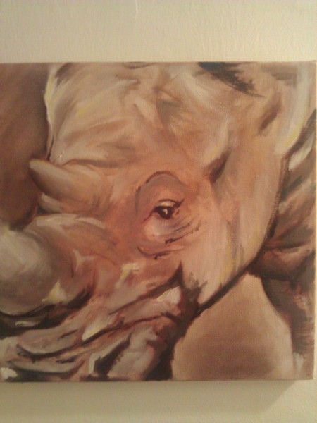
Something exciting that your office has done/is doing for veterinary students this past year: With financial sponsorship from the GHLIT, we are able to provide funding support to all schools for many of these programs. The most recent themes have promoted awareness of rabies, obesity, and vector-borne illness, and the upcoming theme is food safety & food security. Scott's finest piece
Your favorite SCAVMA related experience (EB or not): At the 2012 SAVMA Symposium I was able to meet and interact with the celebrity elephant from Water for Elephants – it was an amazing experience with an enormous and impressive creature.
Something fun about yourself: I am currently working toward a future mastery of the fine art of balloon animal creation. My finest piece is a lion, and I'm trying to improve my "monkey in a tree"
Share this Post | Comments Off Executive Board in SAVMA News