 Eleven Danish veterinary students from the University of Copenhagen visited the University of Florida CVM this past November as part of an International Veterinary Students' Association (IVSA) exchange trip. The UF students will be visiting Copenhagen this May. The IVSA is an international association of which SAVMA is a member.
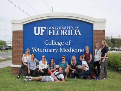
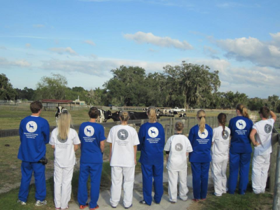
  While the US is an active participant in the IVSA meetings, this is only the second group exchange to take place in the US. During the exchange in Florida, students went to St. Augustine Beach, took a behind the scenes tour at SeaWorld, went alligator "hunting" along Gainesville's nature trails, toured the UF College of Veterinary Medicine and a local animal shelter, visited a brewery, and ate plenty of barbecue at American parties. The American students are looking forward to visiting Copenhagen in May.
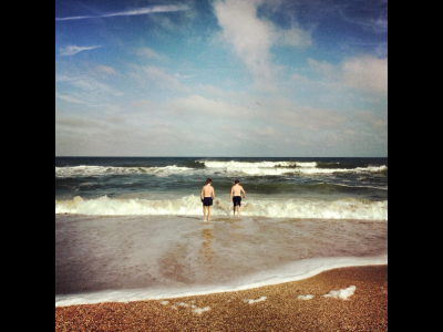

 Share this Post | Comments Off