
Student Case Study: Malignant Oral Melanoma Leads to Intraocular Melanocytic Metastasis in the Canine Patient
Ashley Cubb, Lucien Vallone, Erin Scott, Micheal Deveau, Christian Stocks
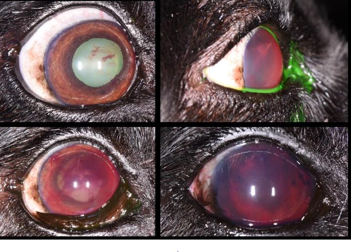
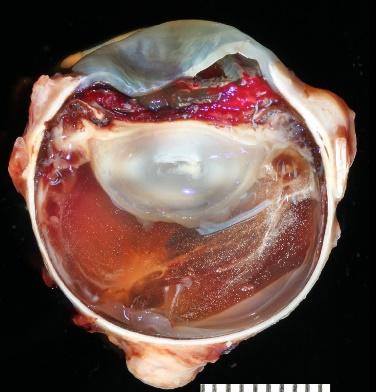
Background: A 12-year-old neutered male Labrador Retriever, with a history of an incompletely excised malignant oral melanoma, presented to the Texas A&M Ophthalmology service with a concern of vision loss. Upon examination bilateral fibrinous uveitis was noted along with secondary glaucoma in the patient’s left eye. Over the next month, uveitis in the left eye continued to progress and hyphema developed. A palliative enucleation was performed on the patient’s left eye and the globe was submitted for histopathology. Objective: Determine the cause and origin of progressive uveitis and hemorrhage in the enucleated eye. Methods: Histopathology of the removed globe and immunohistochemistry using an immunohistochemical stain for melanin-A. Results: Histologic analysis showed a poorly differentiated metastatic mesenchymal neoplasm lining the posterior iris surface, anterior lens capsule, ciliary body surface, and peripheral ventral retina. Immunohistochemistry determined that approximately 80% of the neoplastic cells exhibited strong cytoplasmic positivity for melanin-A, therefore indicating melanocytic origin. Conclusion: Oral melanoma is known for its high metastatic propensity and is one of the most common oral malignancies encountered in canine patients. While primary melanocytic tumors are common ocular neoplasms, secondary ocular melanocytic metastasis as seen in this case have rarely been documented. By observing malignant melanoma patterns of metastasis, development of effective treatment and prevention of this neoplasm could be improved in the future.
 Share this Post | Comments Off