Wednesday
Jan192022

Chats with the Chatfields- EPM

Feeling a bit EPM-ish? No? Perhaps your horse is...
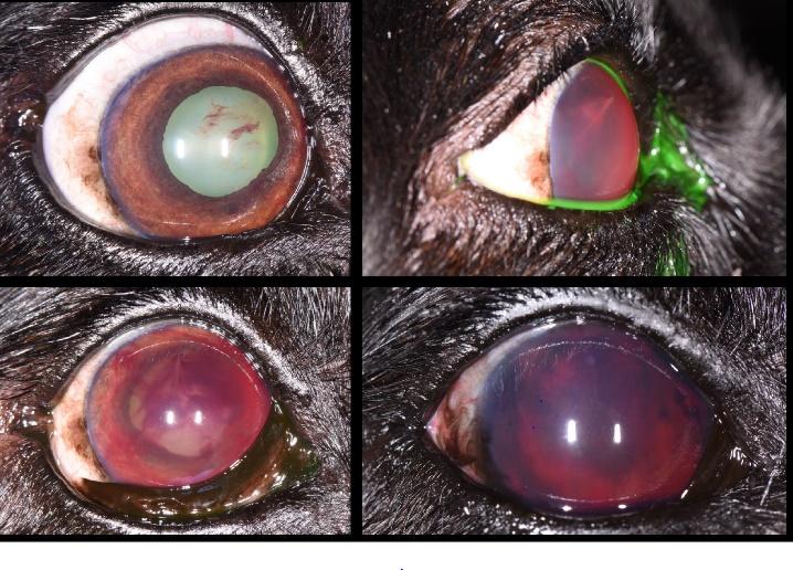
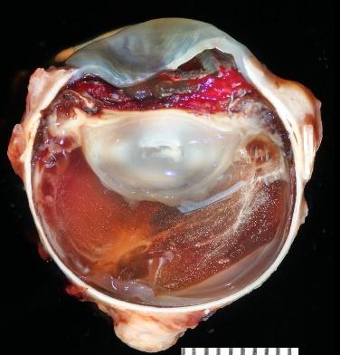
Equine protozoal myelitis (EPM) is a mystery to many in the horse industry - but not to Dr. Rob Franklin!  Dr. Franklin is a board-certified equine internal medicine specialist and has worked with the world's foremost experts on EPM.  Thank goodness he joins Dr. Jen the vet and Dr. Jason Chatfield in the Chat Room to talk all about EPM: the cause, what it looks like in your horse, and what you can do to prevent it!  
Don't worry companion animal friends - Dr. Jen brings in how this is similar to Toxoplasma gondii  (the causative agent of Toxoplasmosis), so there is something in it for everyone!
There's also a bit of V's View from Vet School tucked into this episode at the 12:35 mark! Should you take a break?!  V's got a view on that!
EPM in horses podcast link:
Want more EPM?  Dr. Franklin suggests checking this out: https://onlinelibrary.wiley.com/doi/full/10.1111/jvim.13834
Want to know more about Dr. Franklin?  Check him out here: https://www.fredequine.com/dr-franklin 
SUBSCRIBE to our show on Youtube or on our website: https://chatfieldshow.com
Follow us on instagram @Chatfield_Show
Thanks to our sponsor, FullBucket Veterinary Strength Supplements - the leader in digestive health for horses, dogs, and cats!
V's View is brought to you by the AVMA Trust - Veterinarian inspired coverage protecting you through it all


Tuesday
Jan182022

Photography

Submitted by: Brooke Weinstein, Oregon State University

Friday
Jan142022

Student Case Study: Malignant Oral Melanoma Leads to Intraocular Melanocytic Metastasis in the Canine Patient

Ashley Cubb, Lucien Vallone, Erin Scott, Micheal Deveau, Christian Stocks

Background: A 12-year-old neutered male Labrador Retriever, with a history of an incompletely excised malignant oral melanoma, presented to the Texas A&M Ophthalmology service with a concern of vision loss. Upon examination bilateral fibrinous uveitis was noted along with secondary glaucoma in the patient’s left eye. Over the next month, uveitis in the left eye continued to progress and hyphema developed. A palliative enucleation was performed on the patient’s left eye and the globe was submitted for histopathology.  Objective: Determine the cause and origin of progressive uveitis and hemorrhage in the enucleated eye. Methods: Histopathology of the removed globe and immunohistochemistry using an immunohistochemical stain for melanin-A. Results: Histologic analysis showed a poorly differentiated metastatic mesenchymal neoplasm lining the posterior iris surface, anterior lens capsule, ciliary body surface, and peripheral ventral retina. Immunohistochemistry determined that approximately 80% of the neoplastic cells exhibited strong cytoplasmic positivity for melanin-A, therefore indicating melanocytic origin. Conclusion: Oral melanoma is known for its high metastatic propensity and is one of the most common oral malignancies encountered in canine patients. While primary melanocytic tumors are common ocular neoplasms, secondary ocular melanocytic metastasis as seen in this case have rarely been documented. By observing malignant melanoma patterns of metastasis, development of effective treatment and prevention of this neoplasm could be improved in the future.


Thursday
Jan132022

Calm Before the Storm

By Tayor Curtis, Texas A&M University

Wednesday
Jan122022

Experiences: 1st Place

Semester at the Museum by Lauren Bynum, Texas A&M University


There is nothing more amazing to think about than all the living things that have called Earth home, and to me, nowhere captures the breadth of that quite as well as natural history museums. To see the fossils of creatures that existed before trees did, to be able to stare up through the bones of a dinosaur knowing that it was walking around just a hundred million years earlier—there is nothing like it. One of my favorite parts of attending Boston College for my undergraduate degree was the fact that we received free entry into the Harvard Museum of Natural History, and I took advantage of this free entry many times throughout my four years at BC. During my senior year, I received a dream opportunity – I heard through my animal behavior professor that the Harvard Museum of Natural Science was looking for a new mammalogy intern. I emailed the mammalogy department head right away, and my working interview that next week turned into my first day on the job. 

The mammalogy department was not what I expected. I had anticipated that I would be helping with the upkeep of the specimens displayed to the public in the museum’s Hall of Mammals, but I was led to what seemed like an entire different world that I never knew existed. Separate from the museum is a temperature-regulated library, shelves filled not with books but bones and pelts and horns. The full mammalogy collection is so much more than what is on display in the museum, and researchers visit from all over the world to examine the specimens. One of my favorite frequent visitors of the mammalogy collection was a scientist who was studying the healing of pelvic limb fractures. Every time we ran into each other I was greeted with a smile, a wave of the tibia, fibula, or femur of the day, and an explanation of the type of fracture he had most recently located. Most other visitors were graduate students or researchers studying the evolution of one specific creature, their research made easier by Harvard’s football field sized collection all housed in one giant building. 

My main duty consisted of preparing, labelling, and reorganizing the bovid section, which was rapidly outgrowing the shelf space available to it. Shelving the horns and skulls in particular was a rather difficult game of fossilized Tetris, sometimes due to the weight of the skulls, sometimes their size, and sometimes both. Shocking to no one, it turns out that antelope horns have evolved to be many things, but to be easily shelvable was not one of them. My favorite species of the bovids section was the bison, and I lived for the days where I tracked down the bison bones that needed labelling – the scientific name for the American Bison is Bison bison (and if it’s a Plains Bison, you chuck a third “bison” on the end for the best scientific name of all time: Bison bison bison). Another fun fact – I learned that the scientific name for the black rat is Rattus rattus. I’m a sucker for some repetition. 

I worried a little about whether or not it was wise to take this internship. I love natural history specimens, but it seemed like it may be better for my veterinary school application and my resume to work with animals that were still alive. Ultimately, though, I thought back to the reason why I like natural history museums to begin with: they offer so much understanding about the creatures who walked the Earth before us, along with plenty about the creatures walking the Earth now. With that in mind, it seemed like any chance to learn about animals past or present would lend itself to expanding my knowledge in the veterinary field. And it was true! While I can’t say I memorized anywhere near all the names of the bones in a cow skeleton, I certainly did gain a familiarity with bovine species in general, and also a serious interest in bones which has carried over to my love of veterinary radiology. And on top of that, I got to see all the secrets hiding in the archives—or, as us interns call it, the Harvard Museum of Natural History: Extended Edition.

A picture containing indoor, counter, vegetable  Description automatically generated

A person standing in a museum  Description automatically generated with low confidenceA picture containing indoor  Description automatically generatedA picture containing text, indoor  Description automatically generated